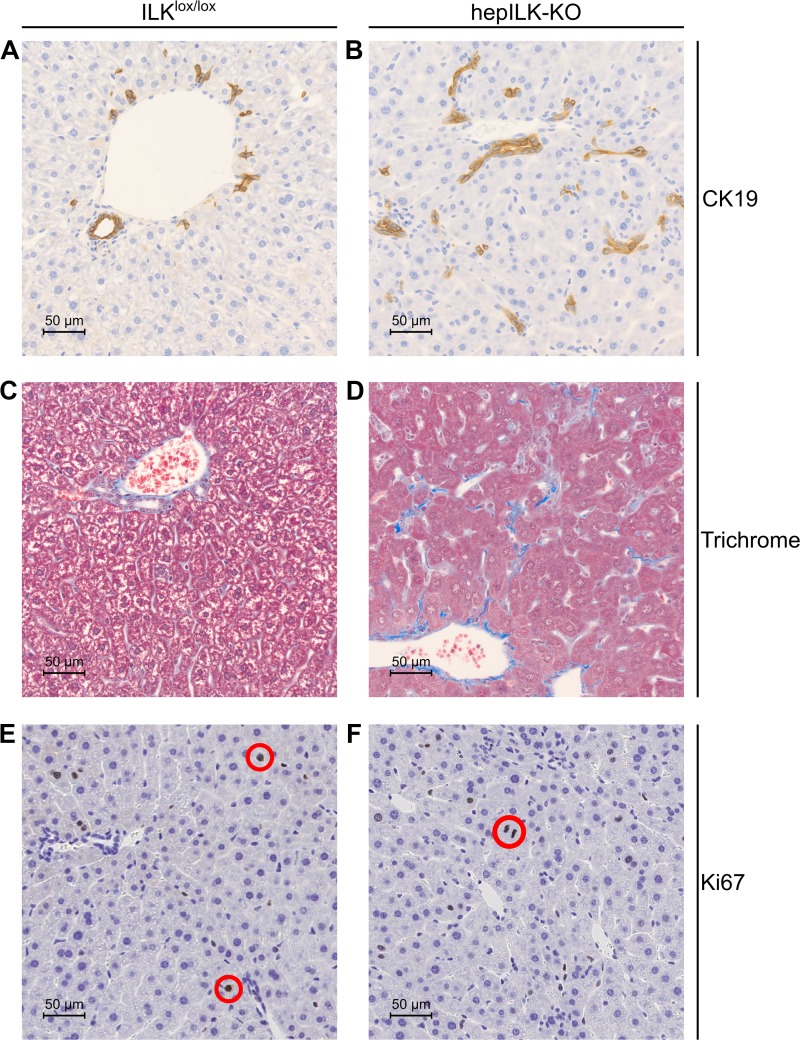
Fig. 2.
A: representative image of immunohistochemical (IHC) staining for CK19 in liver section at ×40 magnification from 6-wk-old integrin-linked kinase (ILK)lox/lox mice. B: representative image of IHC staining for CK19 in liver section at ×40 magnification from 6-wk-old hepILK-knockout (KO) mice. C: representative image of Masson’s trichrome staining in liver section at ×40 magnification from 6-wk-old ILKlox/lox mice. D: representative image of Masson’s trichrome staining in liver section at ×40 magnification from 6-wk-old hepILK-KO mice. E: representative image of IHC staining for Ki-67 in liver section at ×40 magnification from 6-wk-old ILKlox/lox mice. F: representative image of IHC staining for Ki-67 in liver section at ×40 magnification from 6-wk-old hepILK-KO mice. Red circles denote nuclei stained positively for Ki-67 in E and F.

