Fig. 1.
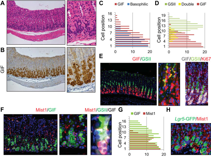
Defining mature chief cells in the mouse stomach. A–C: hematoxylin and eosin (H and E) (A) and gastric intrinsic factor (GIF) (B) staining of wild-type mouse gastric corpus. Sequential sections were stained with the same region. Basophilic cell position on H and E-stained and GIF+ cells are quantified in C. D and E: simultaneous staining for GIF (red) and Griffonia simplicifolia lectin II (GSII) (green) of wild-type mouse corpus (E, left and middle) and cell position quantification of GSII+, GIF+GSII+, and GIF+ cells (D). E, right is triple immunofluorescence for GIF (gray), GSII (green), and Ki67 (red). F, left and middle: immunofluorescence for Mist1 (red) and GIF (green) of wild-type mouse corpus. F, right: immunofluorescence of Mist1 (red), GSII (green), and GIF (gray). G: cell position quantification of GIF+ and Mist1+ cells. H: green fluorescent protein (GFP) (green) and Mist1 (red) staining of Lgr5-DTR-EGFP mice.

