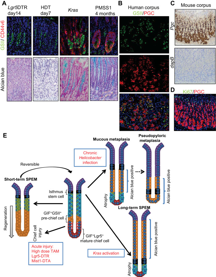
Fig. 8.
Distinct marker expression in acute and chronic metaplasia. A: Griffonia simplicifolia lectin II (GSII) (green)/CD44v6 (red; top) and Alcian blue (bottom) staining of Lgr5-DTR mice 14 days after diphtheria toxin (DT) treatment, wild-type mice 7 days after high-dose tamoxifen (HDT) treatment, Mist1-CreERT; LSL-KrasG12D mice 14 days after tamoxifen treatment, and wild-type mice 4 mo after PMSS1 Helicobacter pylori strain infection. B: pepsinogen C (PGC)/GSII staining in normal human corpus. C: in situ hybridization for Pgc and dapB (negative control) in mouse corpus. D: Ki67 (green)/PGC (red) staining in mouse corpus. E: schematic model of metaplasia development in the mouse corpus gland. SPEM, spasmolytic polypeptide-expressing metaplasia.

