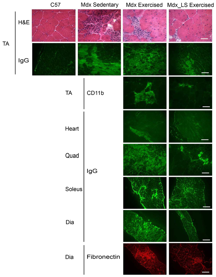
Fig. 1.
Ongoing muscle damage of mice from the Exercised mdx study. Representative images of TA sections from wild-type C57BL/10 (C57), mdx sedentary, and untreated exercised mdx and LS treated exercised mdx mice stained with H&E and IgG. Staining of immune cells for CD11b is also shown for exercised groups. Representative images of IgG staining are shown for heart, quadriceps, soleus and diaphragm sections of untreated and LS treated exercised mdx mice. Fibronectin staining of diaphragm sections shows fibrosis in both untreated and LS treated exercised mdx mice. Bar = 50 μm in CD11b, and H&E images. Bar = 200 μm in IgG and fibronectin images.

