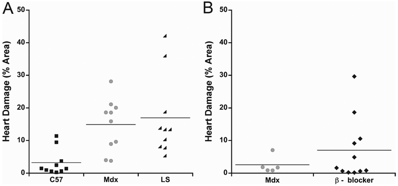
Fig. 4.
Quantification of cardiac damage in isoproterenol treated mdx mice. A) Dot plot showing percentage cross sectional area of IgG immunofluorescence in wild-type C57, untreated mdx, and LS treated mdx after isoproterenol stress; and B) β - blocker treated mdx mice with their untreated mdx cohort after isoproterenol stress.

