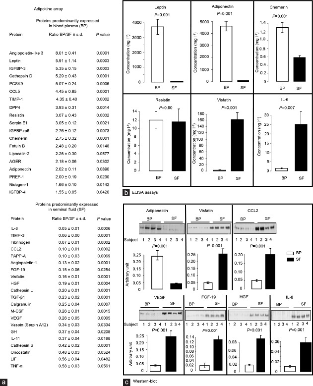
Figure 1.
Expression of adipokines in SF and BP of men of normal weight. (a) Adipokine array data. Proteome Profiler Human Adipokine Array Kit (ARY024 from R&D system, Paris, France) was used as described by the provider to analyze 58 adipokines in BP and SF of seven healthy patients with normal weight and normal semen quality. Data were reported as the ratio between protein expression in BP and in SF (mean ± s.d.). P values were also shown (Student's t-test). Nineteen proteins were higher expressed in BP whereas 21 proteins prevailed in SF. (b) ELISA assays. Plasma and seminal concentrations of leptin, adiponectin, chemerin, resistin, visfatin, and IL-6 were measured using ELISA kits from R&D System (Paris, France, mean ± s.d., n = 25, with serum intra-assay and inter-assay coefficients of variations <8, and for seminal plasma, intra-assay coefficients of variations ≤6% and inter-assay coefficients of variations ≤10%). Data were analyzed by Student's t-test (P values shown). (c) Western blotting. Plasma and seminal concentrations of adiponectin, visfatin, CCL2, VEGF, HGF, and IL-8 quantified by Western blots (mean ± s.d., n = 19). Eighty μg of seminal and BP as determined using bicinchoninic acid assay was denatured in ×5 Laemmli buffer. The samples were then heated at 95°C for 5 min, subjected to electrophoresis on 12% SDS-polyacrylamide gels, and transferred onto nitrocellulose membranes (Schleicher and Schuell, Ecquevilly, France). The membrane was blocked for 30 min in TBS-Tween-milk 5% (v/v) and incubated for 16 h with appropriate primary antibodies at a 1:1000 final dilution. These primary antibodies were obtained from Santa Cruz Biotechnology (Heidelberg, Germany) for VEGF (sc-53462), adiponectin (sc-17044-R), FGF-19 (sc-73984), HGF (sc-1358), CCL2 (sc-32771), and IL-8 (sc-376750) and from Sigma (St Quentin Fallavier, France) for visfatin (V9139). Finally, the blots were incubated for 1 h and 30 min at room temperature with HRP-conjugated anti-rabbit or anti-mouse IgG (dilution 1/5000). The oroteins were detected by enhanced chemiluminescence (Western Lightning Plus-ECL, Perkin Elmer) using a G:Box SynGene (Ozyme) with GeneSnap software (release 7.09.17; Chicago, IL, USA). The signals detected were quantified with the GeneTools software (release 4.01.02; Syngene, Fredrick, MD, USA). The results are expressed as intensity signal in arbitrary units after normalization, allowed by the use of reversible Ponceau staining, as an internal standard. Data were analyzed by Student's t-test (P values shown). s.d.: standard deviation; BP: blood plasma; SF: seminal fluid; HRP: horseradish-peroxidase; IgG: immunoglobulin G; IGFBP-3: insulin-like growth factor binding protein 3; PCSK9: proprotein convertase subtilisin/kexin type 9; CCL5: chemokine (C-C motif) ligand 5; TIMP-1: metallopeptidase inhibitor 1; DPP4: dipeptidyl peptidase-4; AGER: advanced glycosylation end-product specific receptor; PREF-1: preadipocyte factor 1; IL8: interleukin 8; TIMP-3: metallopeptidase inhibitor 3; CCL2: chemokine (C-C motif) ligand 2; PAPP-A: pregnancy-associated plasma protein-A; FGF 19: fibroblast growth factor 19; HGF: hepatocyte growth factor; TGF-β1: transforming growth factor beta 1; M-CSF: macrophage colony-stimulating factor; VEGF: vascular endothelial growth factor; GH: growth hormone; LIF: leukemia inhibitory factor; TNF-α: tumor necrosis factor alpha.

