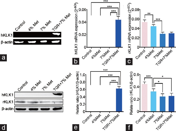
Figure 2.

Verification of the existence and expression of hKLK1 gene in CC of rats. (a) Representative hKLK1 genomic DNA bands in CC through conventional polymerase chain reaction followed by agarose gel electrophoresis. Relative mRNA expression of hKLK1 and rKLK1 genes to β-actin in CC of all four groups by real-time reverse transcriptase polymerase chain reaction: (b) for hKLK1 and (c) for rKLK1. (d) Representative western blot result of hKLK1 and rKLK1 proteins in CC of rats. Western blot results were presented through bar graphs with β-actin as the loading control: (e) for hKLK1 and (f) for rKLK1. Data are shown as mean ± standard deviation (n = 10 per group). *P <0.05, **P <0.01 and ***P < 0.001. Met: ethionine; TGR: transgenic rats; CC: corpus cavernosum; hKLK1: human tissue kallikrein-1; rKLK1: rat tissue kallikrein-1.
