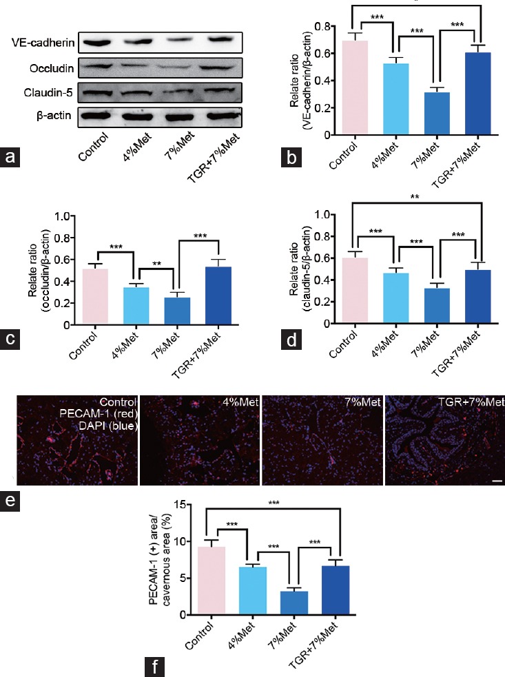
Figure 4.

hKLK1 could preserve EC junction protein expressions and endothelial content in CC of rats with HHcy. (a) Representative western blot results for VE-cadherin, occludin and claudin-5 in CC of rats of all four groups. Expressions of VE-cadherin, occludin and claudin-5 with β-actin as the loading control in all four groups were presented through bar graphs: (b) for VE-cadherin, (c) for occluding, and (d) for claudin-5. (e) Immunofluorescence results of PECAM-1 in rats of all four groups. (f) Ratios of PECAM-1 positive area to cavernous area were presented through bar graphs. Data are expressed as mean ± standard deviation (n = 5 per group). *P < 0.05, **P <0.01 and ***P < 0.001. Scale bars = 100 μm. Met: methionine; hKLK1: human tissue kallikrein-1; CC: corpus cavernosum; HHcy: hyperhomocysteinemia; TGR: transgenic rats; PECAM-1: platelet/endothelial cell adhesion molecule-1; EC: endothelial cell.
