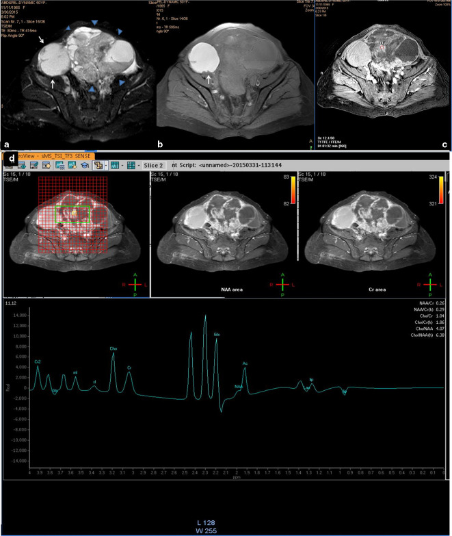
Figure 2. .
A 50-years-old female patient with bilateral ovarian clear cell adenoacarcinoma. (a, b) Axial T 2- and T 1 weighted fat suppressed images that showed bilateral complex masses adnexal/ovarian of mixed intensity; the right-sided mass is the smaller (white arrows) is mainly cystic with few mural-based nodules and the one on the left side displayed thick sepatations and papillary vegetations and wide solid sheets (arrow heads). Both cysts showed complicated content which is most likely haemorrhagic as it showed intense bright signal on the T 1-weighted image. (c) Axial contrast-enhanced image (dynamic series at 1:32 min. post-contrast injection) that showed contrast uptake of the solid component (circle). (d) Multivoxel spectrum with localization on the solid component showed sharp choline signal that present metabolic activity. Note, the multicomponents of the mass presented a complex spectral pattern in the region from 0.5 to 2.5 ppm. The right-sided ovarian mass was false negative and the left-sided one was true positive. MRS,MR spectroscopy; NAA, N-acetyl aspartate

