Figure 3. .
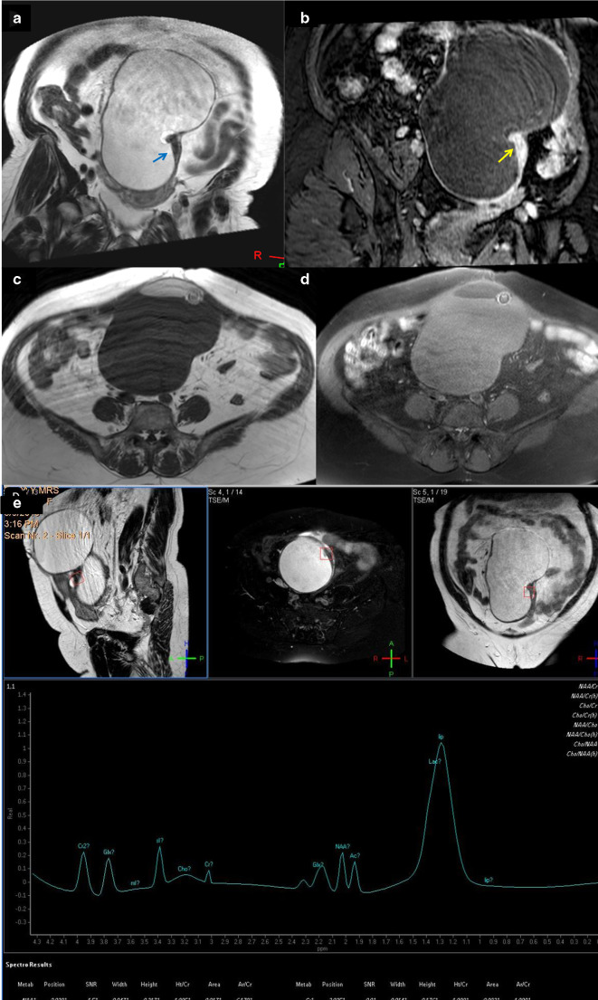
A 62-years-old female patient with immature teratoma with on top squamous cell carcinoma. (a) Coronal T 2-weighted image showing large predominantly cystic complex ovarian mass that showed localized small surface area solid sheet (arrow). (b) Contrast-enhanced coronal image that showed intense contrast uptake of the solid component (arrow). (c, d) Axial T 1- and T 1 fat suppression showed suppression of the bright T 1 signals that suggest fatty elements. (e) Single-voxel MRS, the voxel of interest focused on the solid component and there was large sharp lactate peak, N-acetyl aspartate and acetone signal and muffled choline peak (0.17). N-acetyl aspartate is attributed to the neural elements within the teratoma, the acetone presented reaction due to adherence of the different integrants within the mass. The choline peak was not significant, so it was a false negative, yet when we combined the lactate peak with the choline, malignancy was suggested and the case was considered as true positive. MRS, MR spectroscopy; SNR, signal-to-noise ratio.

