Figure 4. .
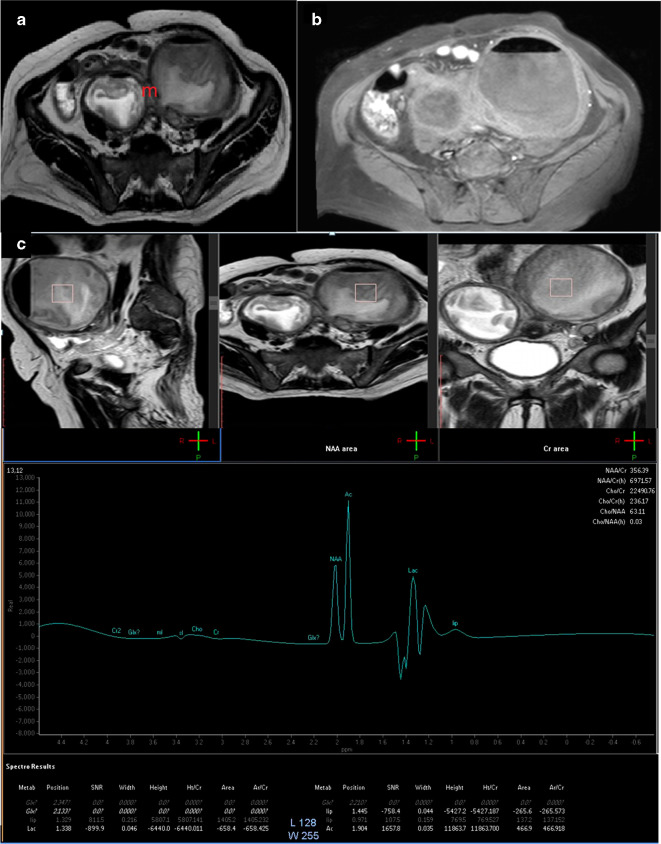
A 30-years-old female patient with bilateral tubo-ovarian abscess. (a) Axial T 2-weighted image showing bilateral cystic ovarian masses (m) that showed thickened walls, the larger on the left side, showed intracytsic indistinct solid sheets. (b) Axial post-contrast injection image showed contrast uptake of the thickened walls yet the intracystic solid component showed no uptake. (c) Single-voxel MRS, showed absence of choline peak. Lactate peak was detected. The presence of N-acetyl aspartate/acetone signal suggests tissue reaction due to turbid/complicated cystic component. The case was true negative. MRS, MR spectroscopy; SNR, signal-to-noise ratio.

