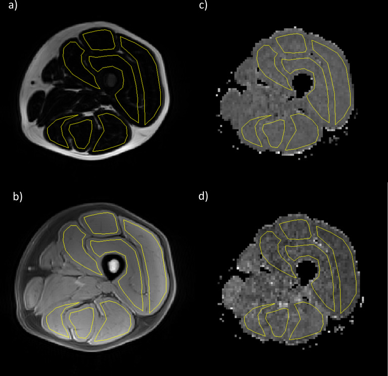
Figure 1.
Example images from a healthy volunteer showing VIBE Dixon (a) fat and (b) water images and STEAM diffusion maps (c) MD and (d) FA. Regions of interest (shown in yellow) were drawn corresponding to the individual muscles of the hamstrings and quadriceps. FA,fractional anisotropy; MD, mean diffusivity; STEAM, stimulated echo acquisition mode; VIBE, volume interpolated breath-hold examination.

