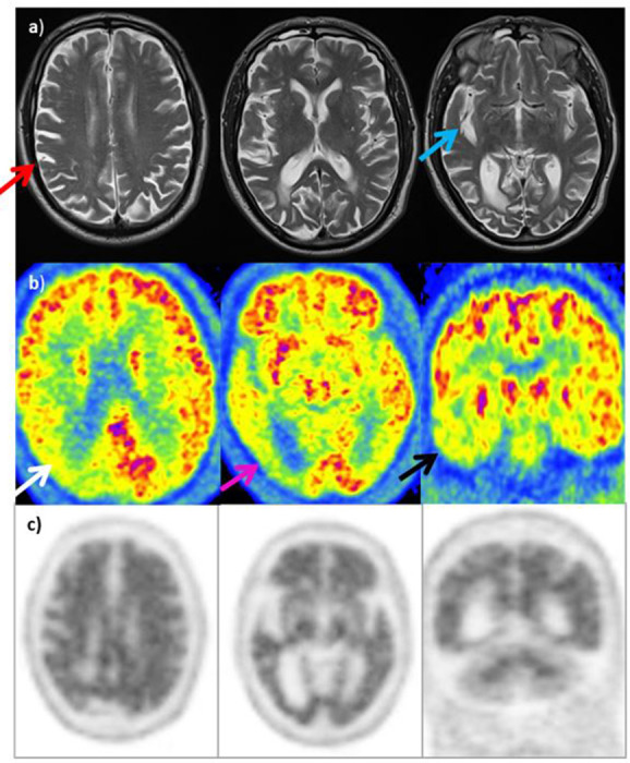
Figure 11. .

MRI (row A): Asymmetric atrophy (right>left) involving right parietal lobe (red arrow) with prominence of right sylvian fissure (blue arrow). Degree of asymmetry atypical for posterior cortical atrophy, but possibly compatible with CBD. 18F-FDG PET (row B): Marked hypometabolism in the right parietal lobe (white arrow), notably posteriorly and in the right occipital lobe (purple arrow). In addition, decreased activity in temporal lobes bilaterally, right>left (black arrow) as well as less marked reduction in activity in the left posterior parietal lobe and occipital lobe. Distribution of hypometabolism suggestive of PCA. Amyloid PET (row C): Loss of grey-white differentitation throughout entire cerebrum in keeping with a positive scan (type A) supporting a clinical diagnosis of AD-type pathology rather than CBD.
