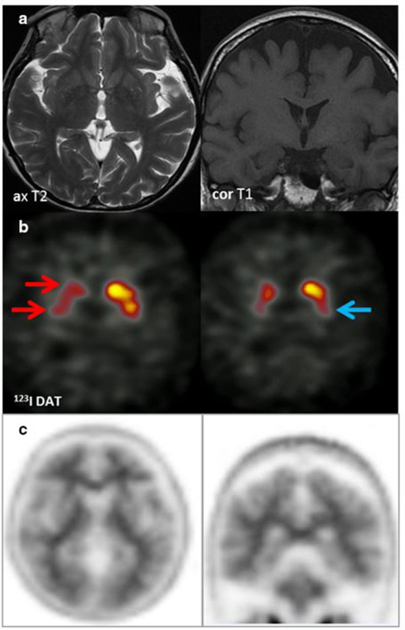
Figure 12. .

MRI (row A): No focal or regional atrophy; hippocampi within normal limits. 123I DaT scan (row B): Severely reduced uptake right caudate and putamen (red arrows) & moderately reduced uptake in the left putamen (blue arrow). Appearances suggestive of DLB. Amyloid PET (row C): Tracer uptake within the cerebral matter similar to that of cerebellar grey matter. Well-defined grey-white matter interface with good definition of the anteroposterior white matter tracts. Characteristic ‘branching-tree’ appearance on coronal PET images, which indicates normal grey-white differentiation in both the cerebrum and cerebellum in keeping with a negative scan indicating absence or sparse levels of amyloid plaque deposition. The functional imaging appearances supported a clinical diagnosis of DLB rather than AD.
