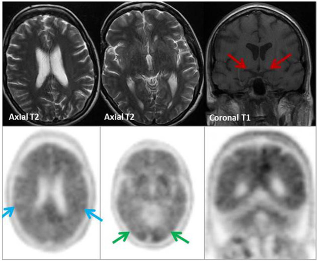
Figure 9. .
MRI (top row): No focal/generalised volume loss. Hippocampal volumes within normal limits (red arrows). 18F-florbetapir (Amyvid™) PET/CT (bottom row): Global loss of grey-white differentiation within the temporal, frontal, parietal (blue arrows), and occipital lobes (green arrows) with ‘tree-in-bloom’ appearance on coronal PET. Appearances are in keeping with a positive scan (type A), supporting a clinical diagnosis of AD.

