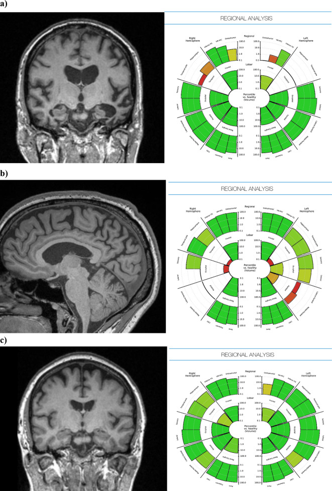
Figure 2. .
Patient-specific anatomical volumes and respective normative data can also be generated by brain region and presented as an easily interpreted and clinically useful graphic report. Examples from patients with (a) established bilateral medial temporal atrophy, (b) posterior cortical atrophy, and (c) healthy appearing brain.

