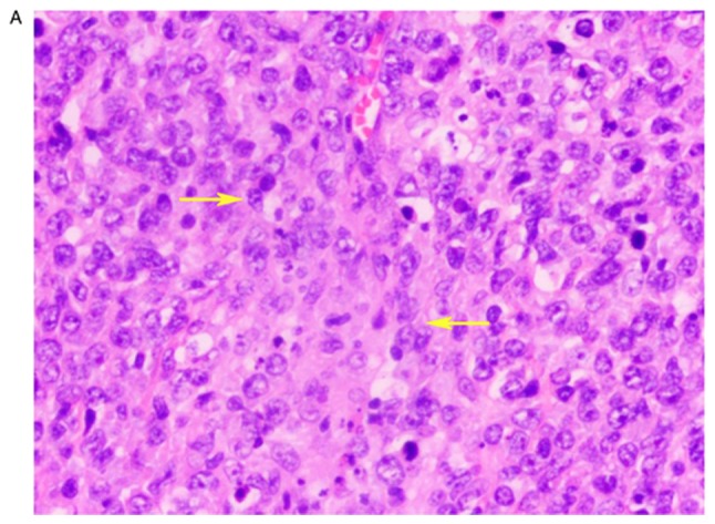
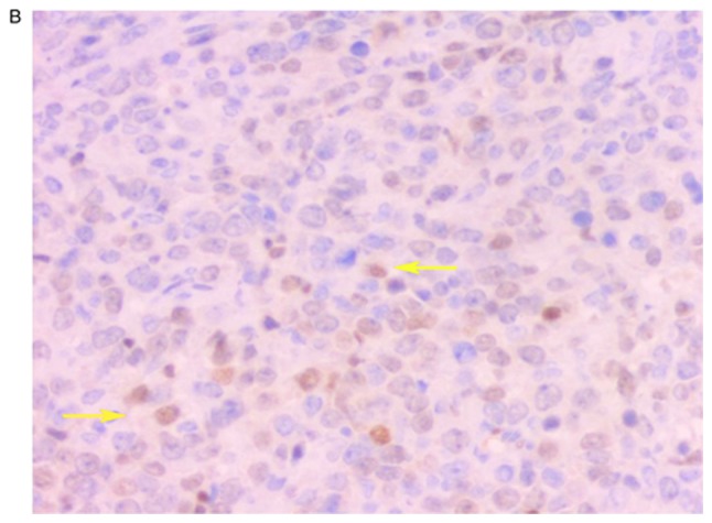
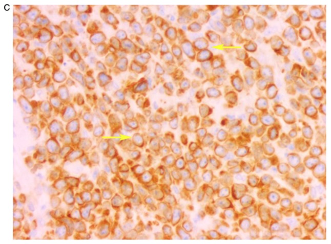
Figure 1.


Case 1. (A) Histological staining of the uterine cervix demonstrating diffuse infiltration of lymphoid cells. These cells were seen in the interstitial tissues. The nucleus was deeply stained with hematoxylin and eosin, and the number of nucleoli was 1–2. Lymphoid tumor cells are indicated (yellow arrows). The mitotic figures were visible. Magnification, ×200. Immunohistochemical staining demonstrating positive expression of (B) proto-oncogene protein (cMyc) in neoplastic cells (yellow arrows). The nucleus was deeply stained and (C) neoplastic cells expressed BCL-2 protein (yellow arrows), with a deeply stained cell membrane. Magnification, ×200.
