Figure 2.
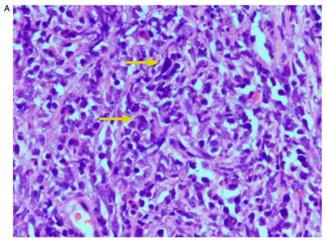
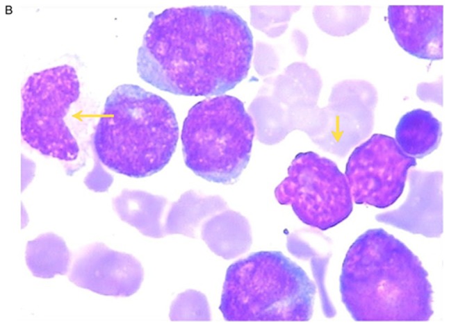
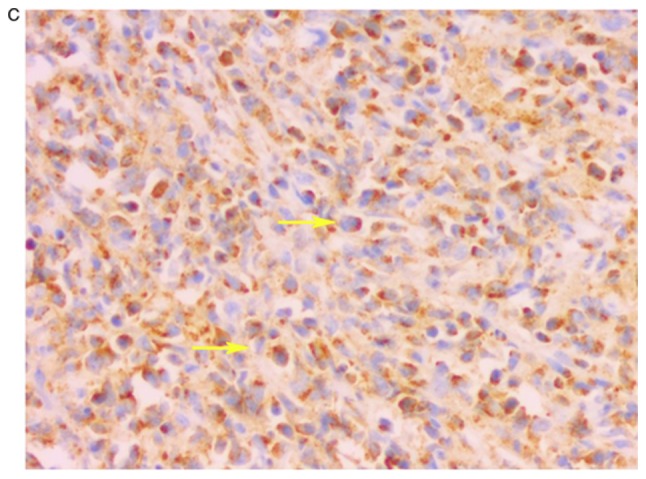
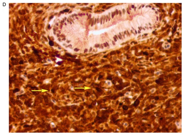
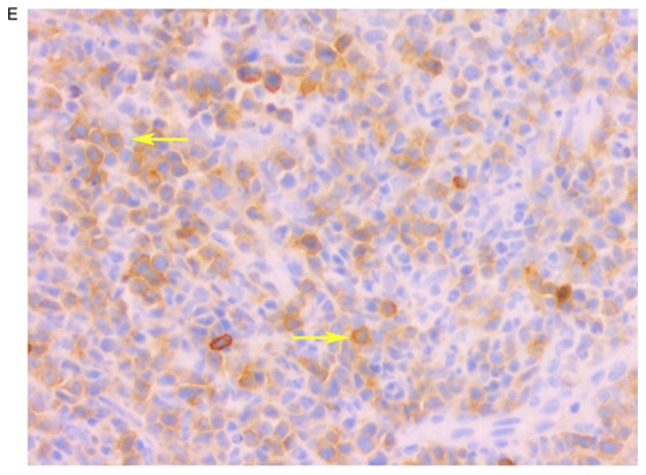
Case 5. (A) Histological staining of the uterine cervix demonstrating neoplastic cells (yellow arrows) were scattered with lymphocytes and inflammatory cells. The nucleus was deeply stained with hematoxylin and eosin, and the nucleolus was not obvious. Magnification, ×200. (B) Bone marrow smear demonstrating myeloblasts (yellow arrows) with large nuclei, deep chromatin and coarse particles. Magnification, ×1,000. (C) Immunohistochemical staining demonstrated neoplastic cells (yellow arrows) with positive expression of myeloperoxidase (MPO) in the cytoplasm, with scattered granular matter. (D) Neoplastic cells had more dark-stained lysozymes (yellow arrows), and the cytoplasm was positive for lysozyme. (E) Neoplastic cells expressed mast/stem cell growth factor receptor (CD117) (yellow arrows), and the cytoplasm and membrane were positive for CD117. Magnification, ×200.
