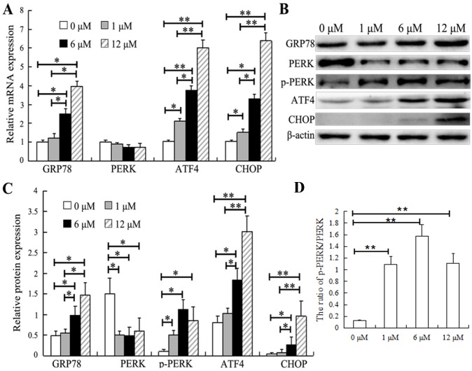
Figure 3.
SAHA induces ER stress in HepG2 cells. (A) HepG2 cells were treated with SAHA at the indicated concentrations for 48 h, and the mRNA levels of endoplasmic reticulum stress-associated genes (GRP78, PERK, ATF4 and CHOP) were analyzed by reverse transcription-quantitative PCR. (B and C) Representative images (one of three experiments) showing the (B) western blot analysis of GRP78, PERK, p-PERK, ATF4 and CHOP protein expression in HepG2 cells exposed to SAHA at the indicated concentrations and (C) their relative protein expression levels. (D) Relative ratio of p-PERK/PERK. N=3. *P<0.05, **P<0.01. SAHA, suberoylanilide hydroxamic acid; GRP78, 78 kDa glucose-regulated protein; ATF4, activating transcription factor 4; CHOP, C/EBP-homologous protein; PERK, PRKR-like endoplasmic reticulum kinase; p, phosphorylated.

