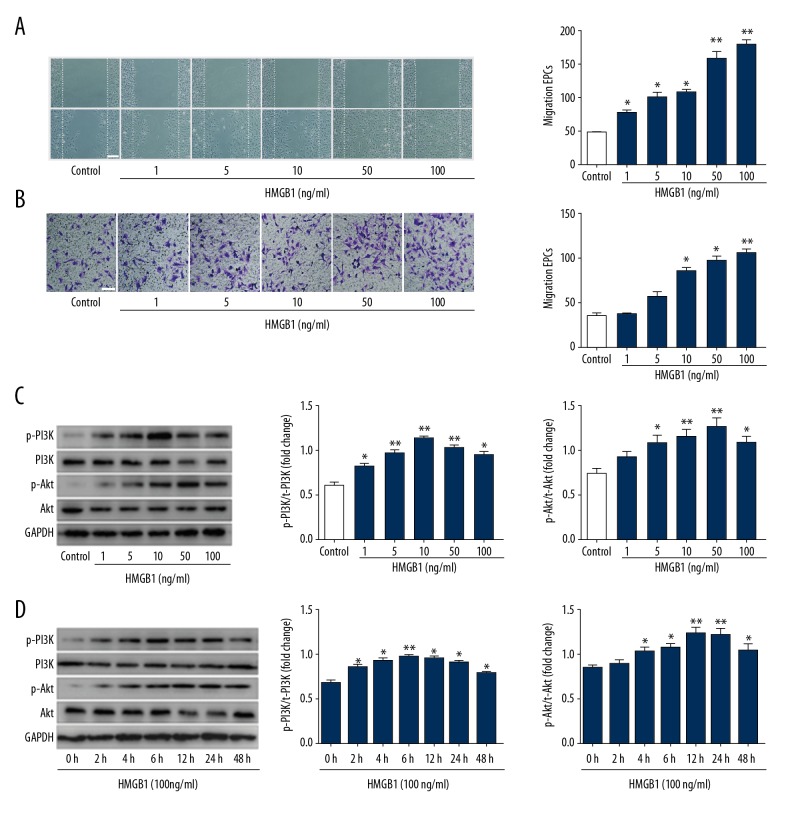
Figure 3.
HMGB1 induced EPCs migration and activated PI3K/Akt/eNOS signal transduction pathways. (A) Wound-healing assay was performed on confluent monolayer EPCs. EPCs were treated with 0–100 ng/ml HMGB1 for 12 h. The wound was monitored and photographed (scale bar=200 μm) 12 h after treatment with HMGB1. Number of EPCs growing into the scratch area was analyzed using Image J software. HMGB1-induced EPC wound healing was concentration-dependent. (B) The migrated EPCs in cell migration assay were stained by crystal violet and photographed 12 h after treatment with HMGB1 (scale bar=100 μm). (C) Western blot was used to measure the interaction of Akt and PI3K protein. (D) EPCs were stimulated with 100 ng/ml HMGB1 for 0–48 h. Expression of p-PI3K and p-Akt was detected using Western blot. The results of Western blot represented as a bar graph. * P<0.05, ** P<0.01, versus control group; # P<0.05, ## P<0.01, versus HMGB1 (100 ng/ml) group.

