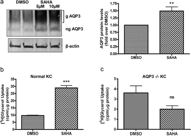
Figure 2. SAHA increases AQP3 levels in situ and AQP3 activity in vitro.
(a) Neonatal mouse skin incubated with medium containing DMSO or SAHA for 24h. A representative (n=3) Western blot for AQP3 levels is shown (left panel). In the right panel, AQP3 levels were normalized to β-actin and expressed as fold over the DMSO-treated group; cumulative results from at least 3 separate skins using DMSO or 5µM SAHA are presented as means±SEM; **p<0.01 versus the DMSO group. (b, c) Keratinocytes from wild-type (b) and AQP3 knockout (c) mice were treated with DMSO or 5µM SAHA for 24h. AQP3 functionality was assessed by [3H]glycerol uptake assay. Please note the change in x-axis (panel b versus c). The data represent means±SEM from 3 independent experiments; ***p<0.001 versus DMSO-treated keratinocytes. g= glycosylated; ng= non-glycosylated.

