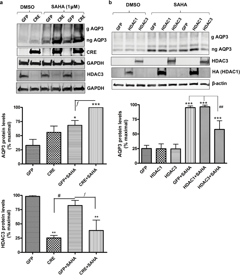
Figure 4. HDAC3 knockdown increases and HDAC3 overexpression decreases AQP3 expression.
Keratinocytes from neonatal floxed HDAC3 mice were infected with Cre- recombinase (CRE)- or control (GFP)-expressing adenovirus for 24h. 57h post-infection the cells were treated with DMSO or 1µM SAHA for 15h. (a) Representative Western blots (upper panel) and quantitation (lower panels), with cumulative values expressed relative to the maximum response as means±SEM from 3 separate experiments. (b) Keratinocytes (wild-type) were infected with adenovirus expressing GFP (control), HDAC3 or HA-tagged HDAC1 for 12h and treated with DMSO or 2.5µM SAHA for 10h. Representative Western blots (upper panel) and quantitation of AQP3 levels (lower panel; n=3) as in (a) is shown. ***p<0.001 versus the control (GFP); fp<0.05, ##p<0.01, #p<0.05 versus the indicated groups. g= glycosylated; ng= non- glycosylated.

