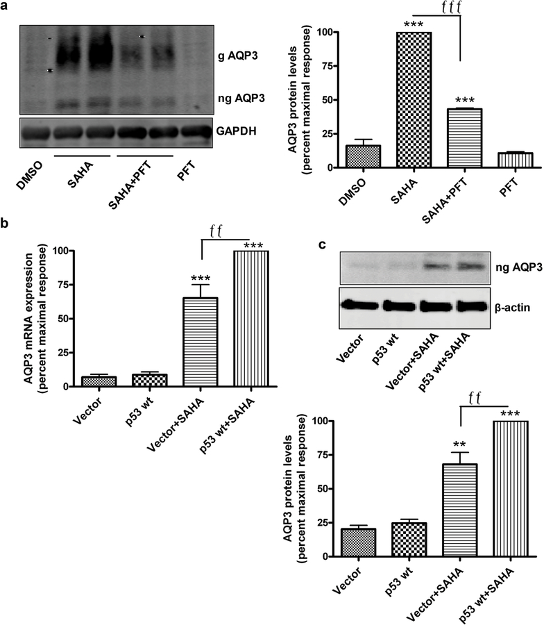
Figure 5. p53 mediates SAHA-increased AQP3 levels in keratinocytes.
(a) Mouse keratinocytes were treated with DMSO or 5µM SAHA in the presence and absence of 30µM pifithrin (PFT) for 24h. Representative Western blot for AQP3 (left panel) and quantitation of 3 experiments (right panel) are shown. AQP3 levels normalized to GAPDH and expressed relative to the maximal response are presented as means±SEM; ***p<0.001 versus DMSO and fffp<0.001 as indicated. (b,c) Mouse keratinocytes were infected with adenovirus expressing wild-type p53 or vector for 12h and treated with 5µM SAHA for 12h. AQP3 mRNA expression analyzed by qPCR (b) and protein levels by Western blots (c, upper panel) with quantitation of 3 experiments are shown (c, lower panel). Results presented as means±SEM are expressed relative to the maximal response. **p<0.01 versus vector, ffp<0.01 as indicated.

