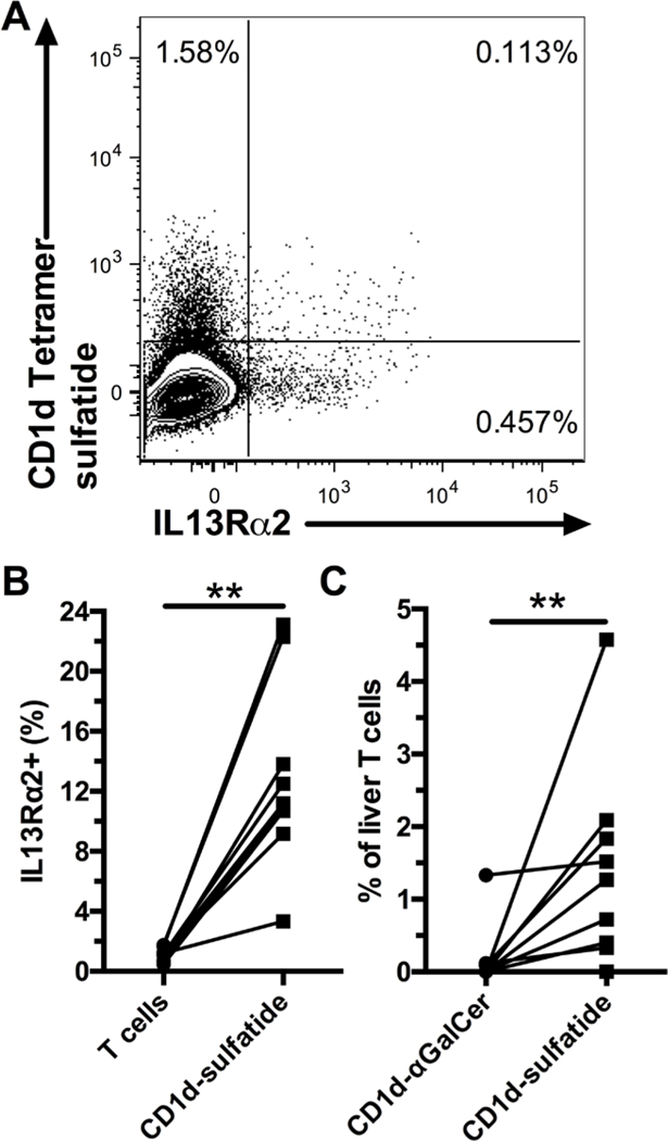
Figure 3. A minority of intrahepatic IL13Rα2+ T cells are type II NKT cells.

Representative staining of liver T cells for IL13Rα2 and CD1d tetramer loaded with sulfatide (A). Frequency of IL13Rα2+ cells within liver T cells and CD1d-sulfatide+ T cells (B, n=9). Frequency of CD1d-αGalCer+ and CD1d-sulfatide+ liver T cells (C, n=9), background staining with unloaded tetramer was subtracted. ** indicates p < 0.01.
