
Figure 3.
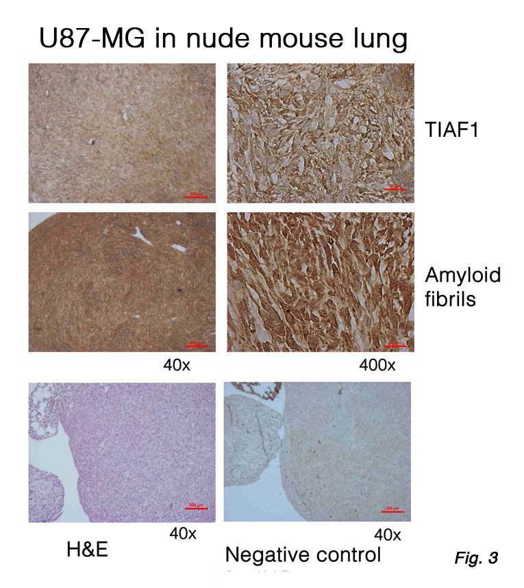
Expression of TIAF1 and amyloid fibrils in U87-MG glioma cells in the lung. U87-MG glioma cells were inoculated in both flanks of nude mice. Two months later, the mice were sacrificed. Shown is the U87-MG cell metastatic to the lung. Each solid tumor lesion has increased expression of TIAF1 and amyloid fibrils, compared to normal mice (data not shown). Both proteins appear to localize intracellularly. A homemade TIAF1 antibody and commercial amyloid fibril antibody were used, as described [34, 35].