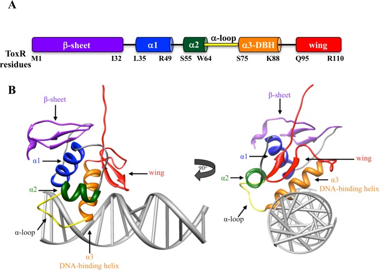
Fig 1. Domain arrangement of ToxR and modeled structure of ToxR on DNA highlighting those domains.
A) Based on homology to other w-HTH proteins [7] the residues defining each domain of ToxR are highlighted. B) A modeled structure of ToxR bound to DNA was generated with the I-TASSER modeling program (http://zhanglab.ccmb.med.umich.edu/I-TASSER/) and the crystal structure of other w-HTH family members [26]. Binding of ToxR to DNA was modeled using the NMR structure of PhoB bound to DNA [25]. Domains were highlighted with the same color scheme used in part A.

