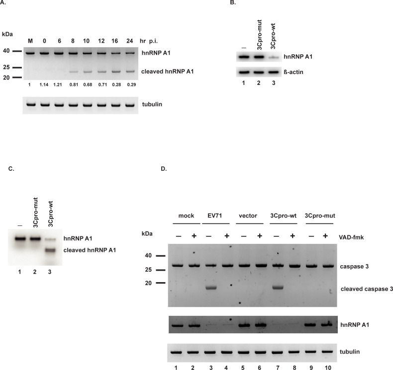
Fig 1. EV71 3Cpro cleaves hnRNP A1.
(A) SF268 cells were infected with EV71 at an moi of 10 or mock infected for 24 hours (lane M). Cell lysates were collected at the indicated time points post-infection. Full-length and truncated hnRNP A1 levels were determined by Western blotting. Full-length hnRNP A1 was quantified and normalized to tubulin, the loading control. The level in mock-infected cells was set = 1.0. Numbers are listed under the blot for hnRNP A1 (B) Purified recombinant wild-type or mutant 3Cpro was added to cell lysates. Following incubation, hnRNP A1 abundance was determined by Western blotting. β-actin: loading control. (C) hnRNP A1 was labeled with [35S]-methionine by in vitro translation. In vitro cleavage assays were performed with wild-type or mutant 3Cpro. Proteins were resolved by 10% SDS-PAGE and detected by phosphorimager. (D) SF268 cells were mock infected or infected with EV71, or transfected with empty vector, or wild-type or mutant 3Cpro expression construct. Pan-caspase inhibitor zVAD-fmk (or DMSO vehicle) was added at a concentration of 50 μM to the indicated cultures for 1–2 days. Levels of caspase-3, cleaved caspase-3, and hnRNP A1 were determined by Western blotting. Tubulin: loading control.

