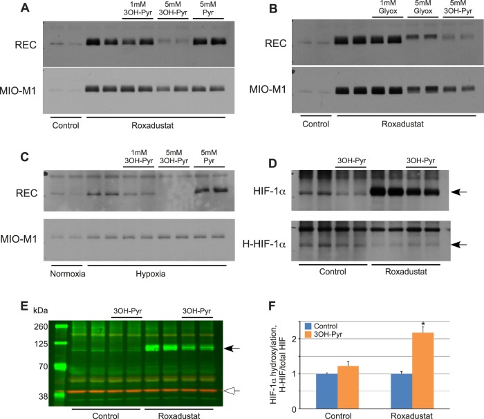
Figure 3.
Effect of 3OH-pyruvate on the cellular levels of HIF-1α and its hydroxylation. (A–C) REC or MIO-M1 cells were first treated with 10 μg mL−1 Roxadustat for 2 hours (A, B) or 2% O2 (hypoxia, [C]) followed by addition of indicated amounts of 3OH-pyruvate, glyoxylate, or pyruvate without changing media (n = 6 for each condition). Cellular proteins were extracted and probed for HIF-1α protein levels by Western blot. (D) REC cells were treated with or without 10 μg mL−1 Roxadustat, 5 mM 3OH-pyruvate in the presence of 10 μM MG-132 for 4 hours (n = 6 for each condition). Cellular proteins were extracted and probed for HIF-1α or hydroxylated HIF-1α (H-HIF) protein levels by Western blot. (E) Representative Western blot image of simultaneous detection of HIF-1α (black arrowhead) and β-actin (white arrowhead). (F) Ratio of hydroxylated/total HIF-1α normalized to β-actin and expressed as fold-change in 3OH-Pyr treated versus untreated REC cells ± SEM obtained by densitometry as described in Methods (n = 4 for each condition). *P < 0.001 versus control.

