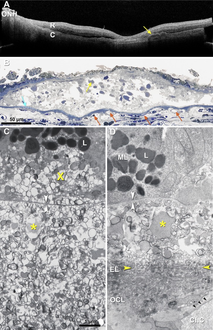
Figure 4.
Soft drusen/basal linear deposit: lipid lakes, no structural collagen/elastin. (A) Ex vivo OCT imaging of a short postmortem donor eye with large soft drusen (arrow, shown in [B]) and central GA, 79-year-old male. R, retina, (C) choroid; ONH, optic nerve head; (B) Large soft druse shown in (A) has numerous lipid pools (arrow) containing esterified cholesterol (refer to Fig. 3C,135 overlaid with dysmorphic RPE and BLamD. The underlying BrM has refractile patches of hydroxyapatite (light blue). Choriocapillaris endothelium ranges from normal to ghosts. Submicrometer section, osmium tannic acid paraphenylenediamine postfixation, toluidine blue stain. (C, D) Soft druse (C) and BLinD ([D] above the yellow arrowheads) from different donors126 contain membranous profiles with electron-dense exteriors and homogeneous and moderately electron-dense interiors, thought to represent partly preserved lipoprotein particles. The same material is found internal (basal mound,8 X) and external to the RPE-BL. Scale bar: 1 μm. Osmium postfixation and transmission electron microscopy. White arrowheads, RPE-BL. Asterisks, lipid lake.

