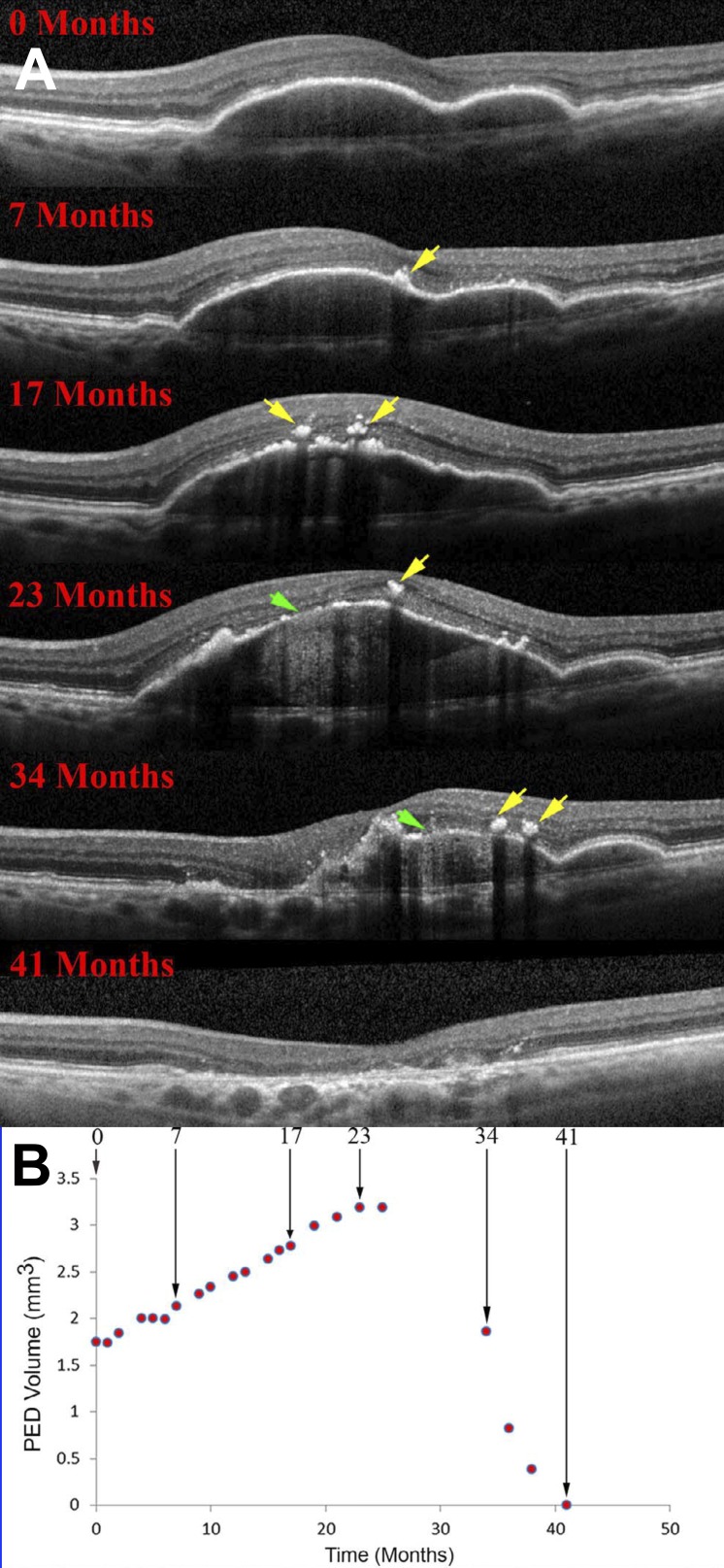
Figure 6.
RPE demise linked to the life cycle of drusenoid pigment epithelial detachment (DPED). (A) Eye-tracked, spectral-domain OCT, in a 72-year-old patient. Intraretinal hyperreflective foci are first noted at 7 months as localized hyperreflective lesions arising from the RPE-BL band (yellow arrows). At 23 months, disruptions to the RPE-BL band (green arrow) with increased light transmission (hypertransmission) to the choroid are evident, followed by reduction in DPED volume until 41 months. (B) DPED volume increased slowly and declined rapidly in this patient. Modified from Balaratnasingam C, Yannuzzi LA, Curcio CA, et al. Associations between retinal pigment epithelium and drusen volume changes during the lifecycle of large drusenoid pigment epithelial detachments. Invest Ophthalmol Vis Sci. 2016;57:5479–5489.

