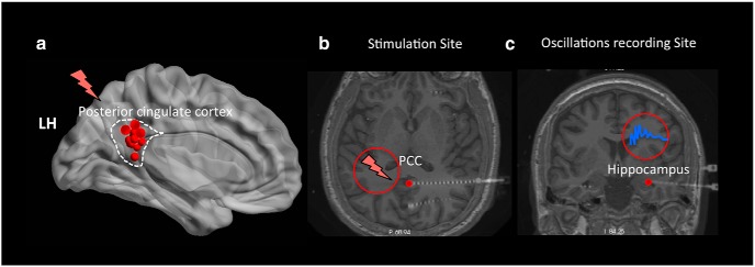
Figure 1.
Electrode placement during a DBS paradigm. a, A summary figure showing the left medial view of the Talairach brain with the locations of stimulated electrodes (red dots) across 16 of the 17 participants who had their stimulation sites in the left PCC. Red lightning bar represents stimulated sites. b, c, Transverse and coronal slices of the sample participant's brain showing the deep insertion of stereo electrodes in the PCC and hippocampus, respectively. DBS was applied to the PCC during the encoding phase in a free recall experiment and neural oscillations were recorded simultaneously from the hippocampus.

