Figure 8.
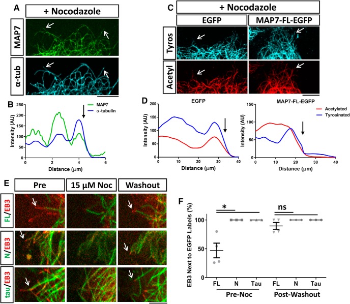
MAP7 prevents nocodazole-induced depolymerization and rescues polymerization after washout. A, Fluorescence images of MAP7-EGFP and α-tubulin staining (arrows) in a region of a COS cell transfected with MAP7-EGFP and treated with nocodazole (6.6 μm). B, Line scans along a single microtubule in (A) reveal that the MAP7 signal terminates at the end (arrow) of the microtubule. C, Fluorescence images of a region of a COS cell transfected with EGFP or MAP7-EGFP and stained for α-tubulin (α-tub) and acetylated-tubulin (actyl). D, Line scans along single microtubules in (C) show that tyrosinated (tyros) microtubules colocalize with acetylated regions after nocodazole treatment. Arrows indicate the end of microtubules. E, Snap shots of COS cell regions expressing MAP7-FL-EGFP, MAP7-N-EGP (green), or tau-EGFP along with EB3-mCherry (red), before (Pre) or during nocodazole treatment (Noc; 15 μm) or after nocodazole washout. Arrows point to EB3-containing growing microtubule ends. F, Quantification of FL, N, and tau-bound microtubules associated with EB3 positive tips pre-nocodazole treatment and post-nocodazole washout (pre; N-FL; p = 0.0297; N-tau; p ≥ 0.999, FL-tau; p = 0.0297; post; N-FL; p = 0.3685; N-tau; p ≥ 0.9999, FL-tau; p = 0.3685, n = 4 for all constructs, Kruskal–Wallis test). Data are reported as mean ± SEM. *p < 0.05; ns, not significant. Scale bars, 5 μm.

