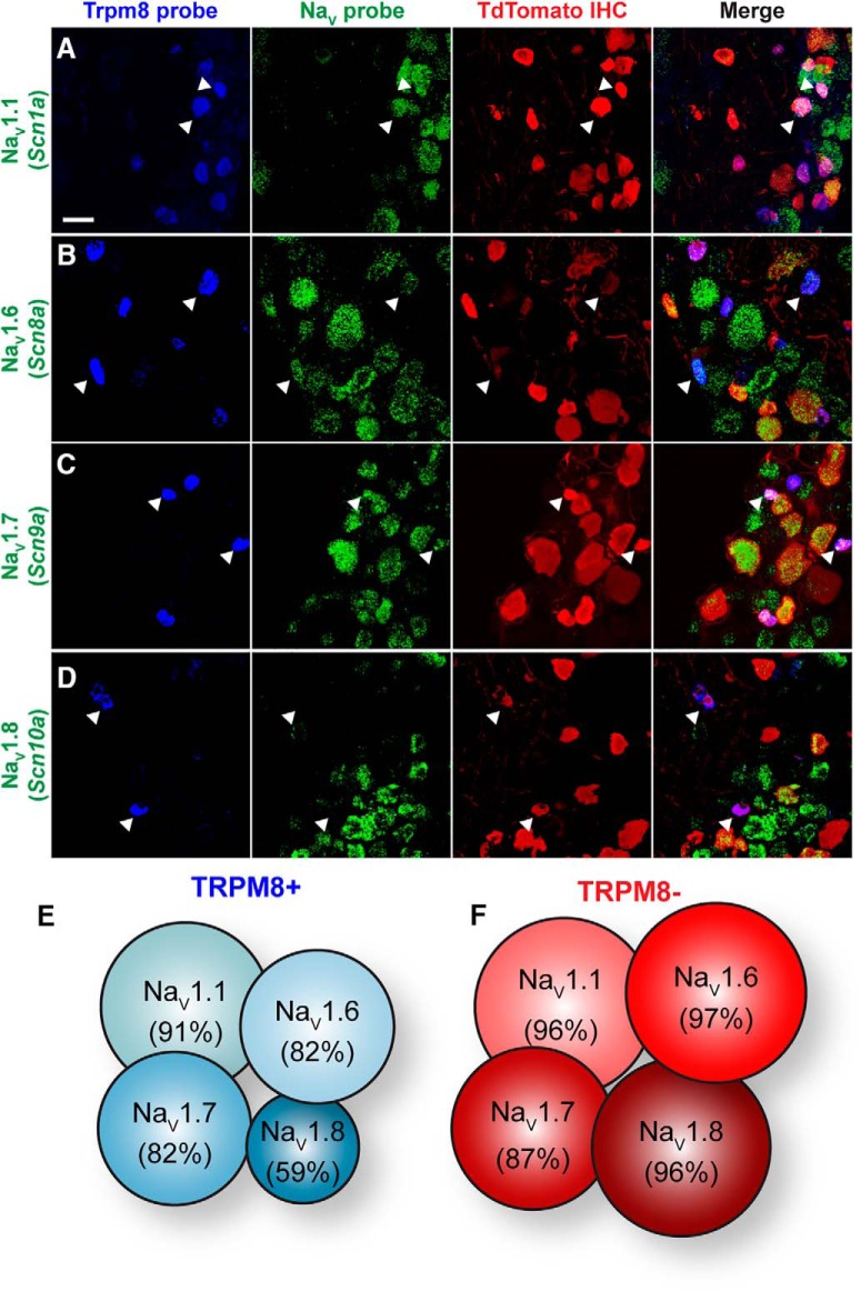
Figure 5.
NaV expression profile of small-diameter Vglut3lineage DRG neurons. A–D, Representative confocal images of single molecule multiplex in situ hybridizations performed on cryosections of adult DRG (25 μm). Images were acquired with a 40×, 1.3 NA oil-immersion objective. Scale bar, 50 μm. Sections were hybridized with probes targeting TRPM8 (Trpm8; blue) and the following voltage-gated sodium channel subunits (green): (A) NaV1.1 (Scn1a), (B) NaV1.6 (Scn8a), (C) NaV1.7 (Scn9a), and (D) NaV1.8 (Scn10a). Sections were stained using immunohistochemistry with anti-dsRED (TdTomato; red) to label Vglut3lineage neurons. White arrowheads indicate representative TRPM8+/NaV+ neurons. E, F, Schematic representation of the percentage of TRPM8+ (E) or TRPM8− (F) small-diameter Vglut3lineage neurons that colabeled for each given NaV subunit.

