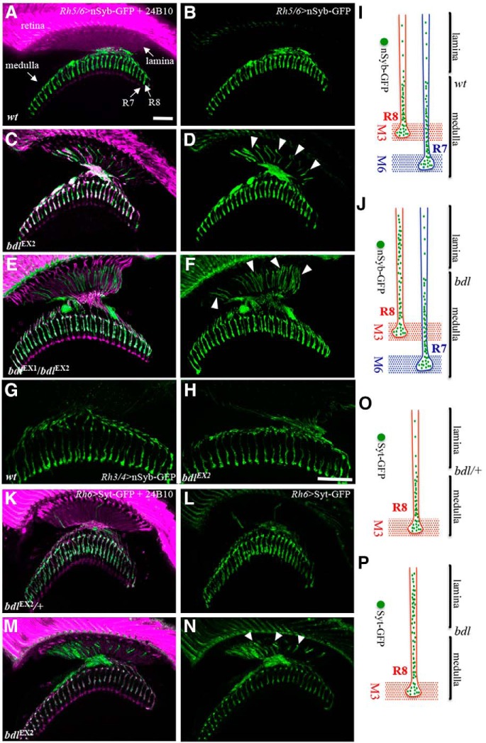
Figure 1.
Many SV components were mislocalized to the proximal portion of R8 axon in bdl mutants. A–D, Frozen sections of adult heads expressing the SV marker nSyb-GFP under control of the R8-specific driver Rh5/6-GAL4 (i.e., Rh5/6>nSyb-GFP), were stained with anti-GFP (green) and MAb24B10 (magenta). MAb24B10 recognizes the cell adhesion molecule Chaoptin expressed in all R-cell axons (Van Vactor et al., 1988). A, In wild-type animals (100%, n = 7), nSyb-GFP staining was predominantly localized to R8 axonal terminals in the medulla region. B, The section in A was visualized with nSyb-GFP staining only. C, In the majority of bdlEX2 homozygous mutant flies examined (6 of 7 animals), strong n-Syb staining was also observed in the proximal portion of R8 axons in the lamina. D, The section in C was visualized with nSyb-GFP staining only. Arrowheads indicate proximal portions of R8 axons with mislocalized nSyb-GFP. E, In all bdlEX1bdlEX2 transheterozygotes examined (n = 6 animals), strong n-Syb staining was observed in the proximal portion of R8 axons in the lamina. F, The section in E was visualized with nSyb-GFP staining only. G, H, Frozen sections of adult heads expressing nSyb-GFP under control of the R7-specific driver Rh3/4-GAL4 (i.e., Rh3/4>nSyb-GFP), were stained with anti-GFP. G, In wild-type (100%, n = 5 animals), n-Syb staining was predominantly localized to R7 axonal terminals in the medulla region. H, In all bdlEX2 homozygous flies examined (100%, n = 5), n-Syb staining was still predominantly localized to R7 axonal terminals in the medulla region. I, J, Schematic illustrations showing the distribution of SV components in R7 and R8 axons in wild-type (I) and bdl mutants (J). K–N, Frozen sections of adult heads expressing another SV marker Syt-GFP under control of the R8-specific driver Rh5/6-GAL4 (i.e., Rh5/6>Syt-GFP), were stained with anti-GFP (green) and MAb24B10 (magenta). K, In bdlEX2/+ heterozygotes (100%, n = 5), Syt-GFP staining was predominantly localized to R8 axonal terminals in the medulla region. L, The section in K was visualized with Syt-GFP staining only. M, In most bdlEX2 homozygous mutants (11 of 13 animals), strong Syt-GFP staining was also observed in the proximal portion of R8 axons in the lamina. N, The section in K was visualized with Syt-GFP staining only. Arrowheads indicate proximal portions of R8 axons with mislocalized Syt-GFP. O, P, Schematic illustrations showing the distribution of SV components in R8 axons labeled with Syt-GFP in heterozygotes (O) and bdl homozygous mutants (P). Scale bar, 20 μm.

