Figure 4.

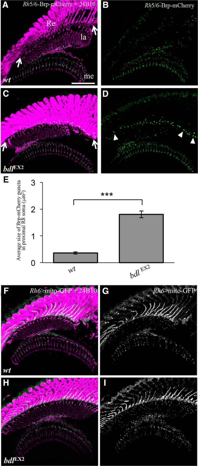
Loss of bdl affected the transport of the AZ protein Brp but not the transport of mitochondria in R8 axons. A–D, Frozen sections of adult heads carrying Rh5/6-Brp-mCherry were double-stained with anti-GFP (green) and MAb24B10 (magenta). A, In wild-type animals (100%, n = 6), Brp-mCherry puncta was predominantly localized to R8 axonal terminals in the medulla region. B, The section in A was visualized with Brp-mCherry staining only. C, In bdlEX2 homozygous mutants (100%, n = 10), abnormal large Brp-mCherry particles were accumulated at the proximal region (arrows) of R8 soma in the retina. D, The section in C was visualized with Brp-mCherry staining only. Arrowheads indicate abnormal large Brp-mCherry particles. E, The size of Brp-mCherry puncta in the proximal region of R8 soma was quantified. Compared with that in wild-type, the size of Brp puncta in the proximal region of R8 soma in bdl mutants showed a significant increase. Student's t test, ***p = 4.5e−07. Error Bars indicate SEM. F–I, Frozen sections of adult heads expressing UAS-mito-GFP under control of the R8-specific driver Rh6-GAL4, were stained with anti-GFP (green) and MAb24B10 (magenta). F, In wild-type (100%, n = 7 animals), mitochondria were detected in both proximal portions of R8 axons in the lamina and R8 axonal terminals in the medulla. G, The section in F was visualized with mito-GFP staining only. H, In bdlEX2 mutants (100%, n = 6 animals), the pattern of mitochondria distribution was similar to that in wild-type. I, The section in H was visualized with mito-GFP staining only. Scale bar, 20 μm.
