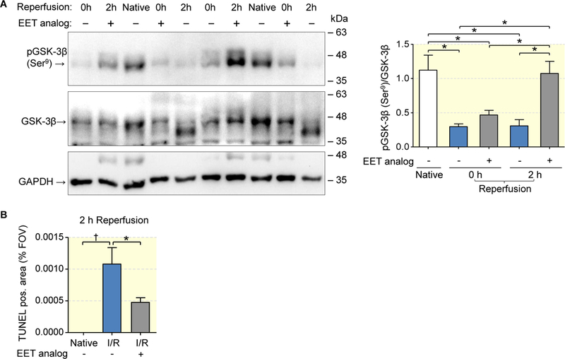
Figure 7: EET analog facilitates the restoration of GSK-3ß phosphorylation and acts anti-apoptotic.

Lower magnitude of dephosphorylation after ischemia and successful rephosphorylation of GSK-3β 2 hours after reperfusion in the EET analog group compared to maintained dephosphorylation of GSK-3β indicative of pro-apoptotic kinase activity in vehicle group (a). Quantitative evaluation of outer medullary sections stained by TUNEL assay confirming lower apoptosis rate in EET analog treated group (b). Data are given as mean ± SEM (n = 6–8 per group in a; n= 4–6 in b). Statistically significant differences were observed as indicated: *(p<0.05); †(p<0.01).
