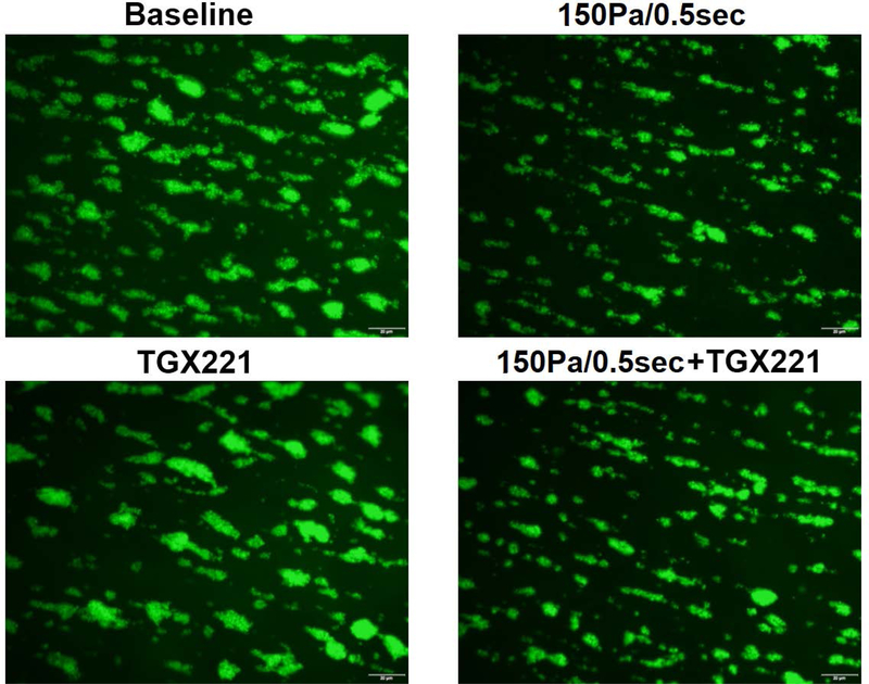
Fig. 5.
The blood samples collected from (1) untreated + unsheared (baseline) condition, (2) TGX221 treated (2.2 μM) + unsheared condition, (3) untreated + sheared (150 Pa/0.5 sec) condition, and (4) TGX221 treated (2.2 μM) + sheared (150 Pa/0.5 sec) condition were perfused through the collagen-coated glass tubes under the physiological shear rate (500 s−1). A) Typical fluorescence images of adherent platelets to collagen; B) Average area coverage (ten images of each glass tube) of adherent platelets to collagen (n=8, *P<0.05).


