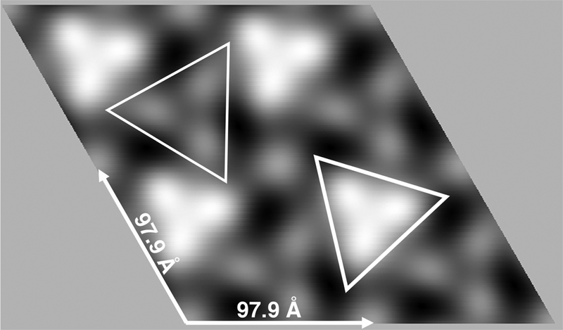
Figure 3. 2D reconstruction of KRAS-GppNHp proteins assembled on membranes.
2D crystals of KRAS-GppNHp proteins assembled on PS-containing membranes were prepared, processed and imaged described in the Methods section. From a set of seven crystals, Fourier transform amplitude and phase data were merged assuming p3 symmetry. The averaged unit cell was a = b = 97.9 ± 5.0 Å, γ = 120.0 + 4.8°. The merge was performed to a resolution of 20Å using reflections of IQ≤6, and the phase residual for the merge was 8.1°, where 0° indicates perfect matching, and 90° indicates random matching. Protein areas appear white, and are viewed perpendicular to the membrane. Bright and faint trimer units respectively are outlined with thick- and thin-lined triangles.

