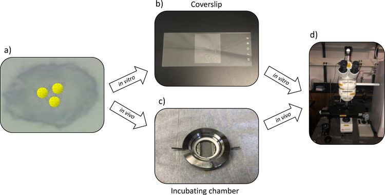
Figure 3).
Workflow to detect αvβ3 integrin on the cell surface with SERS tags. a) Cells were incubated with SERS tags. b) Cells were either fixed onto a coverslip for living cell studies or c) were incubated in a chamber for living cell studies. d) Cell samples, either fixed or living, were Raman mapped.

