
Fig. 3.
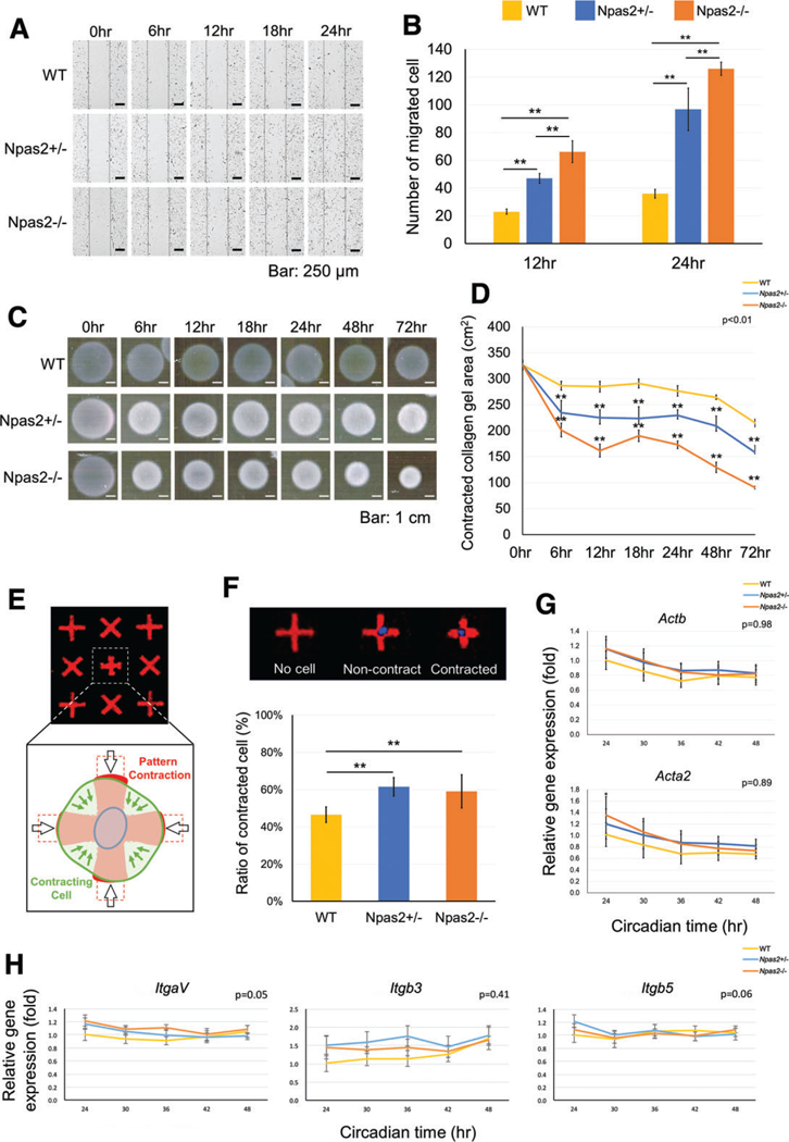
In vitro wound healing experiment using WT, Npas2+/− and Npas2−/− fibroblasts. (A) Images of time-lapse micrographs captured the progressive scratch wound healing assay. The number of migrated cells within the scratched area was significantly larger in the Npas2 KO groups at 6 hr and 12 hr (**P < 0.01). (C) Standardized images of floating collagen gel depicted an increased collagen gel contraction in the Npas2 KO fibroblast groups. (D) The area of collagen gels decreased over time. The gel contraction speed was faster in Npas2 KO fibroblasts (**P < 0.01, significant difference shown only compared with WT). (E) Schematic presentation of the FLECS-based single-cell contraction. (F) The ratio of contracted cells was increased in Npas2 KO fibroblasts. (G) Npas2 KO mutation did not affect the gene expression of β-actin (Actb) and α-SMA (Acta2) in dermal fibroblasts. (H) The steady state gene expression level of integrin subunits αV (ItgaV), β3 (Itgb3), and β5 (Itgb5) in dermal fibroblasts was not affected by Npas2 KO mutation.