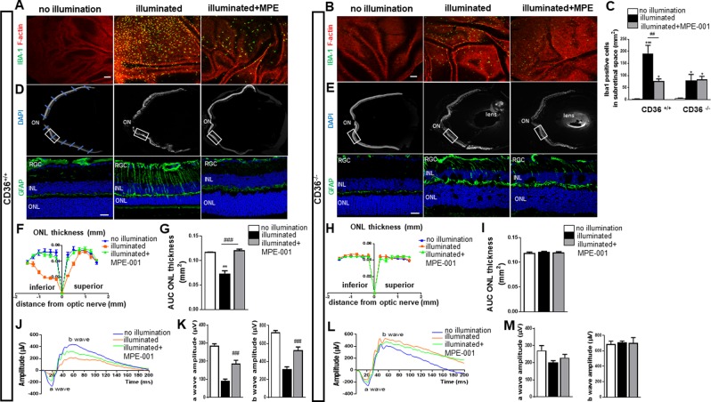
Figure 1.
Azapeptide MPE-001 modulates subretinal MP accumulation and protects against photoreceptors degeneration and function. (A–M) CD36+/+ (n = 6 mice/group) and CD36−/− mice (n = 3 mice/group) were illuminated for 5 days with 6000 lux blue light and subcutaneously injected with 289 nmol/kg per day of MPE-001, starting at 24 h following blue-light exposure for a total of 7 consecutive days. (A,B) F‐actin of RPE cells was counterstained with rhodamine phalloidin (red), showing IBA-1+ MPs (green) in the subretinal space of central retina from CD36+/+ and CD36−/− mice. Scale bar: 25 μm. (c) IBA-1+ cell quantification in the subretinal space. (D,E) Upper panels show representative cryosections with nuclear staining (DAPI) of central retina with the optic nerve (ON) from CD36+/+ or CD36−/− mice. The blue lines indicate the location of measurements of the ONL thickness on each side of the optic nerve (ON). Lower panels show representative images of central retina (12X magnification of white square) stained with GFAP (green). (F,H) Spider graphs of the ONL thickness measured at defined distances of the ON. (G,I) Bar graph showing ONL AUC from CD36+/+(F,G) and CD36−/− mice (H,I). (J–M) Representative scotopic ERG responses from CD36+/+ (J) and CD36−/− mice (l). Quantification of a and b wave amplitudes ERG from CD36+/+ (K) and CD36−/− mice (M) at a light intensity of 3.0 cd*s/m². In C,G,K,M one-way ANOVA test with Newman-Keuls post-test for multiple comparison was performed. *P < 0.05, **P < 0.01 and ***P < 0.001 vs no illumination group. ##P < 0.01 and ###P < 0.001 vs illuminated group. Data are shown as mean ± S.E.M. ONL: Outer Nuclear Layer, INL: Inner Nuclear Layer, GCL: Ganglion Cell Layer.

