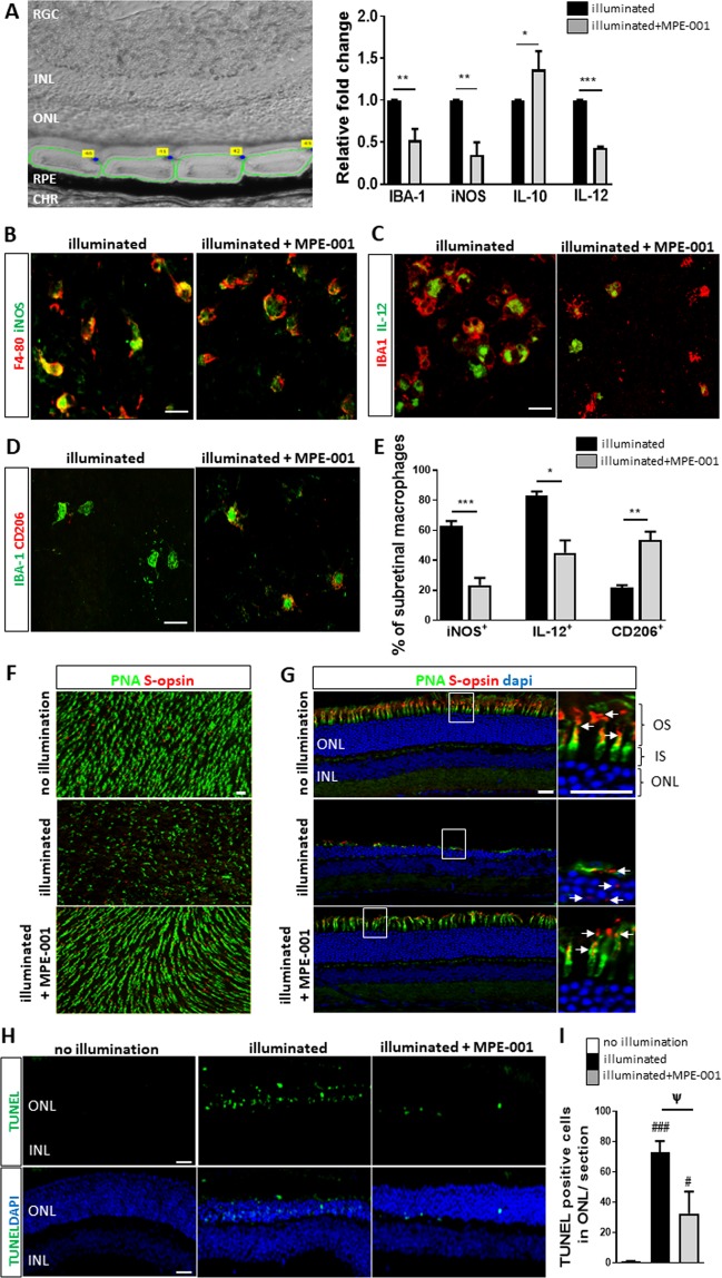
Figure 2.
Azapeptide MPE-001 regulates inflammatory profile of subretinal MPs and reduces photoreceptor degeneration. (A–H) CD36+/+ mice were illuminated for 5 days with blue light. Subcutaneous injections of 289 nmol/kg MPE-001 were administered after 1 day illumination and pursued daily for 7 consecutive days. (A) Upper panel: area of retinal cryosections were microdissected between ONL and RPE and visualized with green circles. Lower panel: bar graphs of IBA-1, iNOS, IL-6, IL-10 and IL-12 mRNA expression levels in microdissected retinal cryosections. Cytokine analysis were normalized to 18 s rRNA. (B) Subretinal MPs stained for F4/80 (red) and iNOS (green) on RPE flat mounts as assessed by confocal microscopy. (C) Subretinal MPs stained with IBA-1 (red) and IL-12 (green) on RPE flat mounts as assessed by confocal microscopy. (D) Subretinal MPs stained with anti-IBA-1 (green) and anti-CD206 (red) antibodies on RPE flat mounts as assessed by confocal microscopy. Scale bar: 15 μm. (E) Percentage of subretinal MPs (IBA-1+ or F4/80+) expressing INOS, IL-12 or CD206. (F) Confocal microscopy of neuroretinal flat mounts (photoreceptors side) and (G) retina cryosections from illuminated CD36+/+ mice treated or not with MPE-001 stained with fluorescein PNA (green) and anti-S-opsin (red) antibody; nuclei were counterstained with dapi (blue). Magnifications of white square show length of cone outer segment with S-opsin distribution. Scale bar: 10 μm. (H) Retinal cryosections stained with TUNEL (green). Nuclear layers were stained with DAPI (blue). Scale bar: 25 μm. (I) Percentage of TUNEL+ cells in ONL cryosections of the retina. In (A and E) unpaired t-test was performed. *P < 0.05, **P < 0.01 and ***P < 0.001 vs illuminated group (n = 3-4 mice/group). In I, one-way ANOVA test with Newman-Keuls for multiple comparison was performed. #P < 0.05 and ###P < 0.001 vs no illumination group. Ψ P < 0.05 vs illuminated group (n = 3-4 mice/group). Data are shown as mean ± S.E.M. RGC: Retinal Ganglion Cell. INL: Inner Nuclear Layer. ONL: Outer Nuclear Layer. RPE: Retinal Pigment Epithelium. CHR: Choroid.

