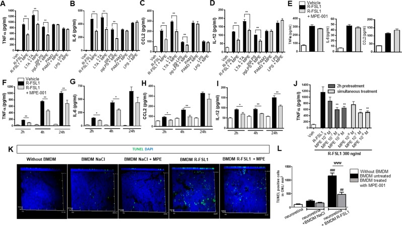
Figure 3.
Selective inhibitory effect of CD36 ligand on TLR2-mediated pro-inflammatory cytokine secretion by MPs and ensued mitigation of photoreceptor apoptosis. (A–D) Pro-inflammatory cytokines TNFα, CCL2, IL-6 and IL-12 concentrations in supernatants of WT peritoneal MPs stimulated with selective TLR2/6 heterodimer agonist (300 ng/ml R-FSL1, 1 µg/ml LTA), TLR2/4 agonist (1 µg/ml pgLPS), TLR2/1 agonist (100 ng/ml PAM3CSK4) and TLR4 agonist (100 ng/ml LPS) for 4 h in the presence of 10−7 M MPE-001 or vehicle. (E) TNFα, IL-6 and CCL2 secretion in supernatants of peritoneal MPs from CD36−/− mice treated with 10−7 M MPE-001 or vehicle, stimulation with 300 ng/ml R-FSL1 for 4 h. Data in A–E are representative of 5-6 independent experiments (n = 3-4/group). (F–I) Time-dependent release of TNFα, IL-6, CCL-2 and IL-12 secretion from WT peritoneal MPs treated or not with 10−7 M MPE-001 following stimulation with R-FSL1 for 2, 4 and 24 h (n = 4/group). (J) Dose-dependent inhibition of TNFα secretion in WT peritoneal MPs after 2 h pretreatment or simultaneous treatment with MPE-001 (10−8, 10−7 and 10−6 M) followed by 4 h stimulation with 300 ng/ml R-FSL1; (n = 8/group). (K) Confocal microscopy of flat mounts with z-stack projections of TUNEL (green) stained neuroretinal flat mounts incubated without or with BMDM stimulated with R-FSL1 or vehicle and treated or not with MPE-001. Nuclei are counterstained with DAPI (blue). (L) Numbers of TUNEL positive cells/mm2 in the ONL of neuroretinal explants incubated or not with monocytes in the different conditions (n = 3-4 eye/group). In (A–I) unpaired t-test was performed. *P < 0.05; **P < 0.01. In J and L one-way ANOVA test with Newman-Keuls for multiple comparison was performed. *P < 0.05; **P < 0.01. ##P < 0.01 and ###P < 0.001 vs neuroretina (without BMDM), ψψψ P < 0.001 vs BMDM untreated. Data are shown as mean ± S.E.M. RPE: Retinal Pigment Epithelium. CHR: Choroid.

