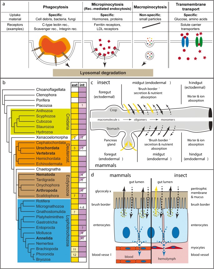
Fig. 1.
The molecular, cellular and phylogenetic basis of extracellular and intracellular digestion. a The basic molecular and cellular principles of nutrient ‘absorption’ by phagocytosis, receptor-mediated endocytosis, macropinocytosis or transmembrane transport. b The phylogenetic distribution of extracellular (‘ext’, yellow) and intracellular (‘int’, purple) digestion activities. White, empty boxes: absence of activity. Half boxes: partial absence of phagocytosis or of intracellular digestion in certain subgroups. Light colours represent minor roles of respective mode. Comments: (1) Uptake of ferritin suggests micropinocytosis; phagocytosis may be present in fibre cells, where an ‘immunity role’ is probable. (2) Xenoturbella: probably only macropinocytosis present (Israelsson 2008). (3) Secretion of lysosomal enzymes in Ciona intestinalis (Thomas 1970) and no phagocytosis, but only pinocytosis of HRP in Oikopleura (Cima et al. 2002). (4) Some nematodes show pinocytosis, but no phagocytosis. (5) Pinocytosis shown but no phagocytosis. (6) Pycnogonids, arachnids and crustaceans have both extra- and intracellular digestion. All other arthropod groups (with some exceptions, such as blood-sucking insects) lack intracellular digestion. (7) Present in Priapulida; in Kinorhyncha, Loricifera: extracellular digestion is likely, considering the presence of gland and zymogen cells; intracellular digestion is unstudied. (9) Cephalopoda present only pinocytosis. (10) Extracellular digestion dominates in most annelids, but intracellular digestion plays a role in leeches. In some polychaetes, phagocytic coelomocytes appear to invade the gut epithelium. (11) Minor role for extracellular digestion; only carbohydrase activity found. (12) Minor role for extracellular digestion. c Comparison between the insect (upper half) and mammal guts (lower half), and their secretory and absorptive capacities. Yellow arrows: secretion of polysaccharidase (e.g. Amylase) and endopeptidase (e.g. Trypsin). Black arrows: secretion of oligo- and disaccharidases, and oligo- and dipeptidases. Dashed arrows: Apical (black) and baso-lateral (blue) transmembrane transport of amino acids and monomeric carbohydrates (e.g. glucose, fructose).d Schematic representations comparing vertebrate and insect enterocytes. Colours as in (c). b is based on following, non-exhaustive list of references: Choanoflagellates (Dayel and King 2014), Ctenophora (Bumann and Puls 1997; Hernandez-Nicaise 1991), Porifera (Imsiecke 1993; Leys and Eerkes-Medrano 2006; Weissenfels 1982; Willenz and Van De Vyver 1982), Placozoa (Grell and Ruthmann 1991; Smith et al. 2014), Cnidaria (Arai 1997; Bouillon et al. 2006; Van-Praët 1985), Xenacoelomorpha (Israelsson 2008; Markosova 1986; Pedersen 1964), Cephalochordata (Biuw and Hulting 1971), Urochordata (Thomas 1970; Yonge 1937), Vertebrata (Karasov and Hume 1997), Hemichordata (Bridges and Woodwick 1994), Echinodermata (Tokin and Filimonova 1977), Chaetognatha (Arnaud et al. 1996), Nematoda (Clokey and Jacobson 1986; Riley 1973; Wright 1991), Tardigrada (Biserova and Mustafina 2015; Dewel et al. 1993; Yonge 1937), Onychophora (Heatley 1936; Manton 1937; Storch and Ruhberg 1993), Arthropoda (Ceccaldi 1989; Fahrenbach and Arango 2007; Filimonova 2008; Miguel-Aliaga et al. 2018; Wägele et al. 1981), Scalidophora (Kristensen 1991; Kristensen and Higgins 1991; Storch 1991; Storch et al. 1989), Rotifera (Wurdak 1987; Yonge 1937), Micrognathozoa (Møbjerg Kristensen and Funch 2000; Yonge 1937), Gnathostomulida (Lammert 1991),Platyhelminthes (Antoniazzi and Silveira 1996; Bowen et al. 1974; Jennings 1968; Ruppert et al. 2004), Gastrotricha (Ruppert 1991; Ruppert et al. 2004; Teuchert 1977), Entoprocta (Morton 1960; Ruppert et al. 2004), Mollusca (Boucaud-Camou and Yim 1980; Lobo-da-Cunha 2000; Owen 1974; Yonge 1937), Annelida (Jennings and Van Der Lande 1967; Jeuniaux 1969; Michel et al. 1984; Yonge 1937), Nemertea (Ruppert et al. 2004), Brachiopoda (Morton 1960; Steele-Petrovic 1976; Yonge 1937), Phoronida (Vandermeulen and Reid 1969), Bryozoa (Yonge 1937)

