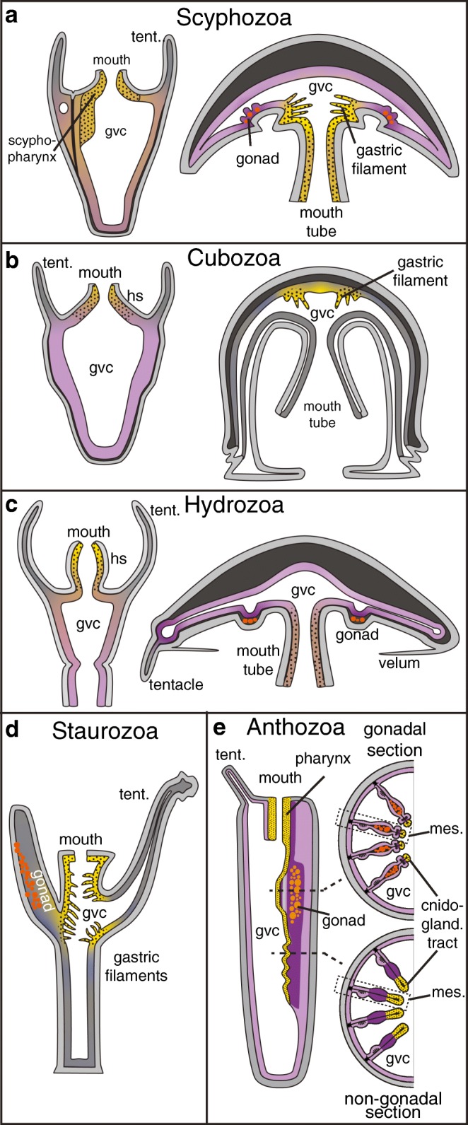
Fig. 3.

a–d The distribution of tissues with extra- or intracellular digestion in scyphozoan (a), cubozoan (b), hydrozoan (c), staurozoan (d) and anthozoan (e) cnidarians. Dotted yellow: exclusively exocrine; purple: exclusively phagocytic/pinocytic; brown: mixed tissues. Grey: no exocrine, phagocytic or pinocytic activity described. Boxed regions in (e) are magnified in Fig. 4a and b. In staurozoans and cubomedusae, no data is available on phagocytic or pinocytic tissues or cell types. Gvc: gastrovascular cavity; hs: hypostome; mes: mesentery; tent: tentacle
