
Fig. 1.
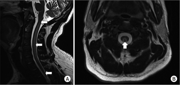
In case 1, the cervical spine T2-weighted magnetic resonance imaging showed an inverted Vshaped hyperintensity (arrow) in the bilateral dorsal column of cervical spinal cord: (A) sagittal view and (B) axial view at C2 level.
Official websites use .gov
A
.gov website belongs to an official
government organization in the United States.
Secure .gov websites use HTTPS
A lock (
) or https:// means you've safely
connected to the .gov website. Share sensitive
information only on official, secure websites.
In case 1, the cervical spine T2-weighted magnetic resonance imaging showed an inverted Vshaped hyperintensity (arrow) in the bilateral dorsal column of cervical spinal cord: (A) sagittal view and (B) axial view at C2 level.