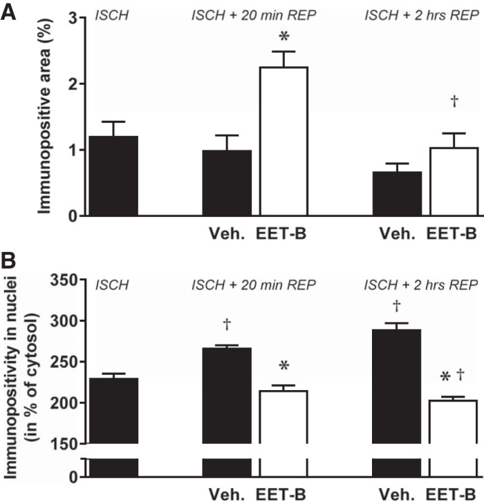
Fig. 8.

Hypoxia-inducible factor (HIF)-1α tissue immunopositivity (A) and prolyl-4-hydroxylase domain protein 3 (PHD3) nuclear positivity (B) in the nonischemic septum assessed at the end of ischemia (ISCH) and after 20 min and 2 h of reperfusion (REP) in rats administrated vehicle (Veh) or the epoxyeicosatrienoic acid (EET) analog EET-B (2.5 mg/kg) 5 min before reperfusion. Values are means ± SE. For HIF-1α, 4 different sections (six images in each) of 3 hearts/group were analyzed; for PHD3, 25 nuclei from 4 different sections of 3 hearts/group were analyzed. *P < 0.05 vs. vehicle; †P < 0.05 vs. ischemia.
