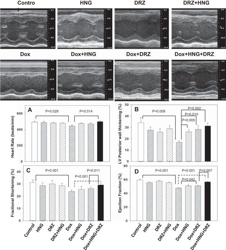
Fig. 2.
Representative M-mode echocardiography photo images of mouse hearts from different treatment groups. A−D: heart rate (A), left ventricle (LV) posterior wall thickening (B), fractional shortening (C), and ejection fraction (D) in control, synthetic humanin analog (HNG) alone-, dexrazoxane (DRZ) alone-, DRZ + HNG-, doxorubicin (Dox) alone, Dox + HNG-, Dox + DRZ-, and Dox + HNG + DRZ-treated mice.

