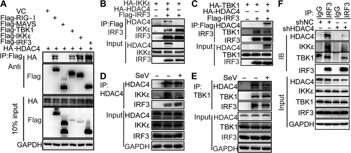
Figure 4.
HDAC4 interacts with TBK1, IKKε, and IRF3. (A) IP and IB analyses of lysates of HEK293T cells overexpressing vector (VC) or plasmids encoding Flag-tagged RIG-I, MAVS, TBK1, IKKε, and IRF3, plus HA-tagged HDAC4, probed with anti-HA or anti-Flag. Bottom, IB analysis of lysates without IP (10% of input). (B and C) IP and IB analyses of lysates of HEK293T cells overexpressing plasmids encoding HA-tagged IKKε or HA-tagged TBK1, HA-tagged HDAC4, and Flag-tagged IRF3, probed with various combinations of anti-HA or anti-Flag. (D and E) Immunoassay of lysates of HEK293T cells (5 × 10⁶) left uninfected or infected for 8 h with SeV, followed by IP analysis with immunoglobulin G (IgG), as a control (first lane), or with antibody to HDAC4 or TBK1 and IB analysis with antibodies to IKKε and IRF3 (D) or HDAC4 and IRF3 (E). Bottom, IB analysis of the samples above (input) without IP. (F) The endogenous association of TBK1, IKKε, and IRF3 in the presence or absence of HDAC4. IP analysis with anti-IRF3 and IB analysis with anti-IRF3, anti-TBK1, anti-IKKε, anti-HDAC4, or anti-β-actin of HEK293T cells infected with SeV for 6 h. Data are representative of three independent experiments.

