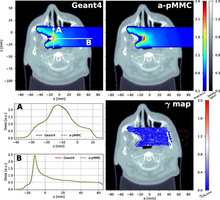
Fig. 8.
Results of the patient CT case. Transversal cut of the head and neck patient CT showing dose color washes for a mono-energetic proton 4×4 cm2 broad beam of 100 MeV calculated with Geant4 (top left) and the adaptive pMMC (top right). Dose profiles as indicated by the white arrows are shown (bottom left) and the result of a 3D-Gamma evaluation with a 1% (global) and 1 mm criterion (20% lower cutoff) is presented for the corresponding slice (bottom right)

